
Electron Microscopy and Spectroscopy
BEG e-MAGE Lab
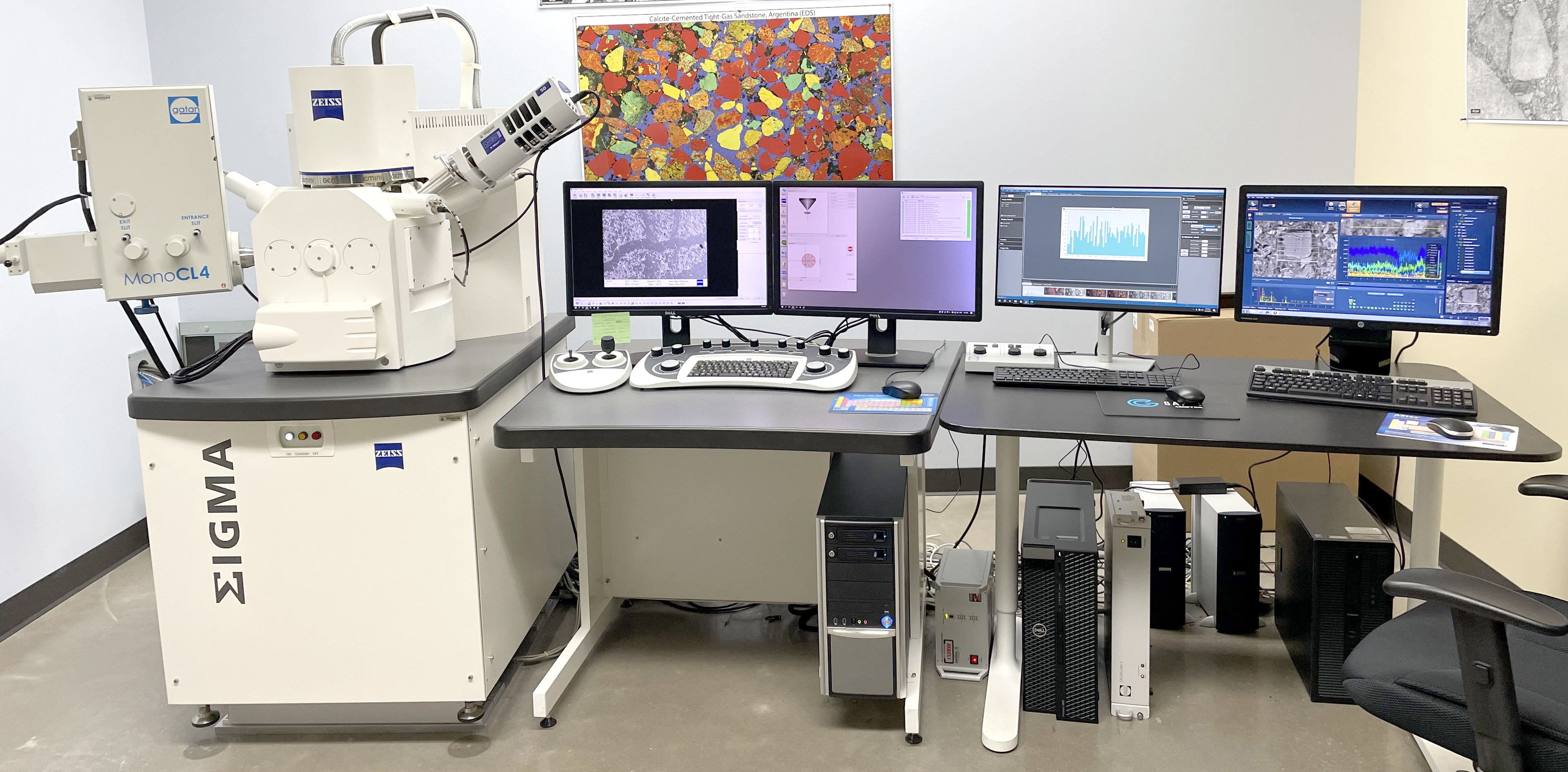
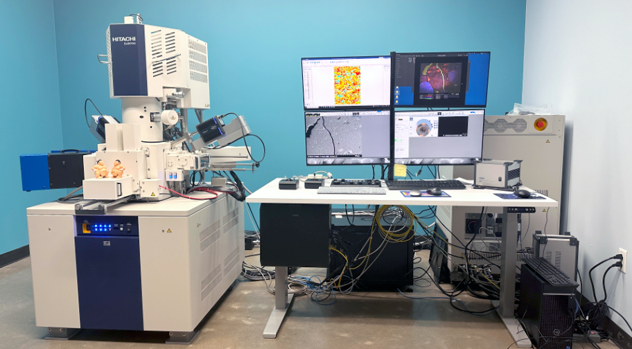
The BEG’s advanced Electron Microscopic Analysis in Geology and Engineering (e-MAGE) Laboratory specializes in chemical and textural microanalyses of natural and engineered samples. The SEM facility is equipped with state-of-the-art imaging, analytical, and sample preparation capabilities.
Equipment Includes:
Specialized Imaging Technique: SEM-Cathodoluminescence
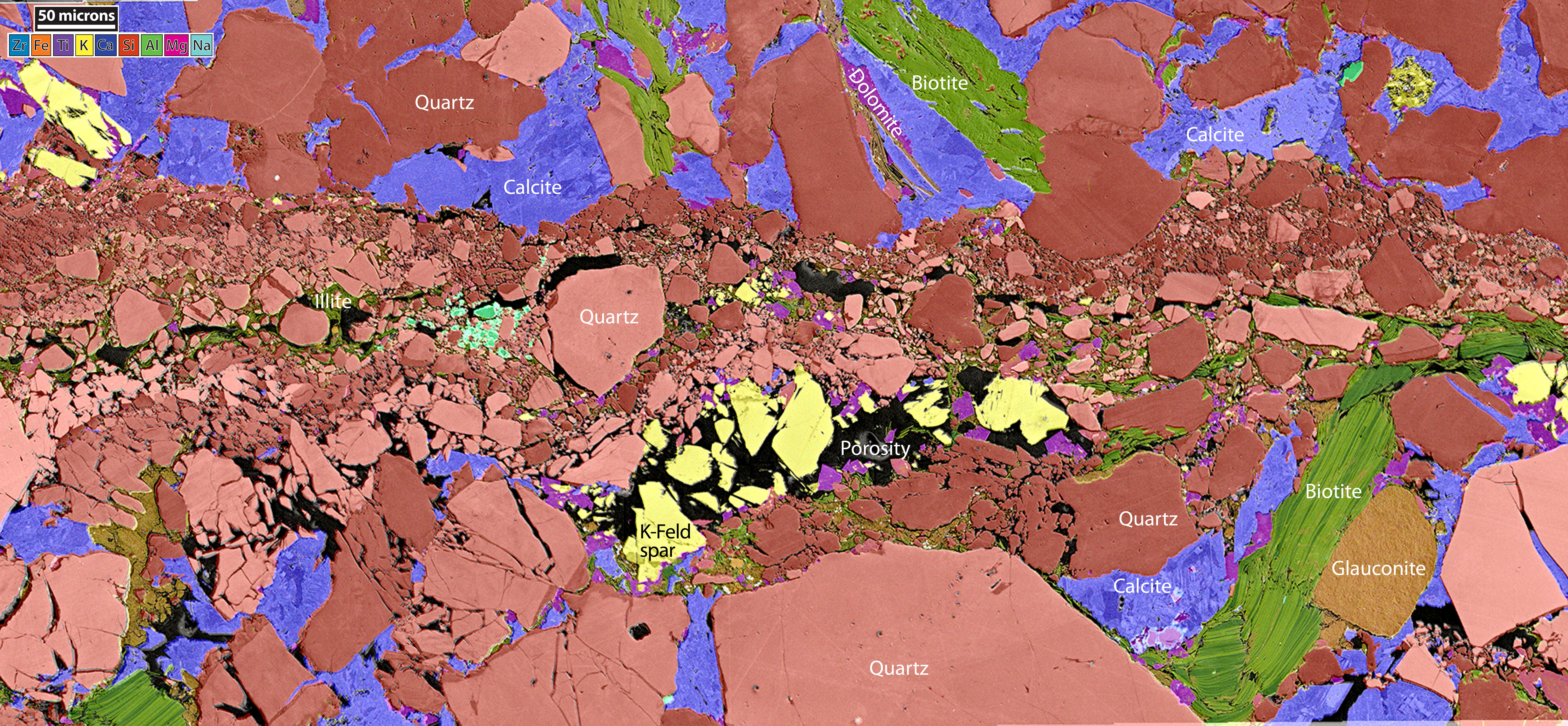
SEM-cathodoluminescence (SEM-CL) is the emission of photons of characteristic wavelengths from a material under high-energy electron bombardment as a function of composition, trace elements, and lattice structure defects. SEM-CL provides information unavailable through other known imaging techniques including detrital grains versus cements, different generations and textures of cements, deformation mechanisms and relationships, and types of porosity including nano- and secondary.
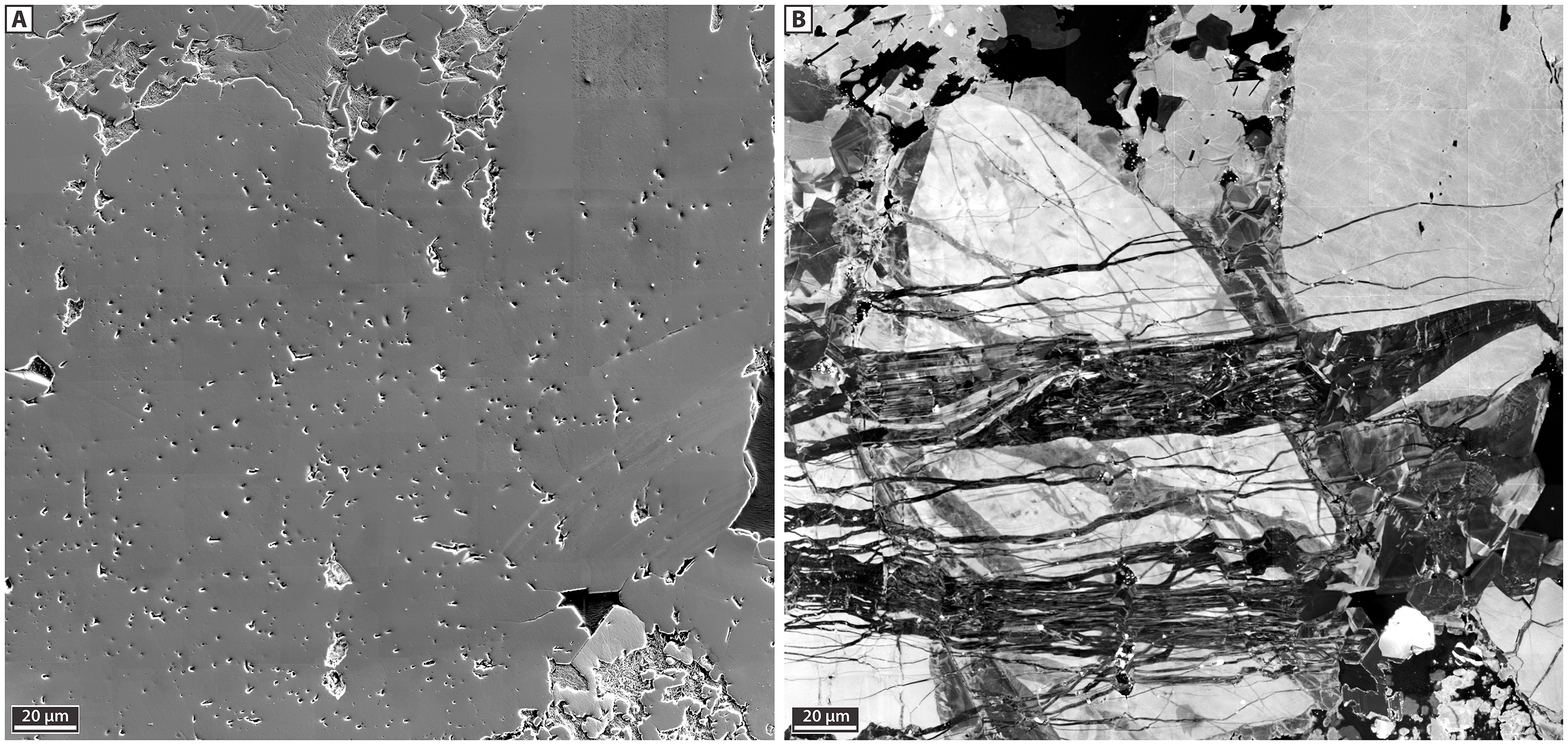
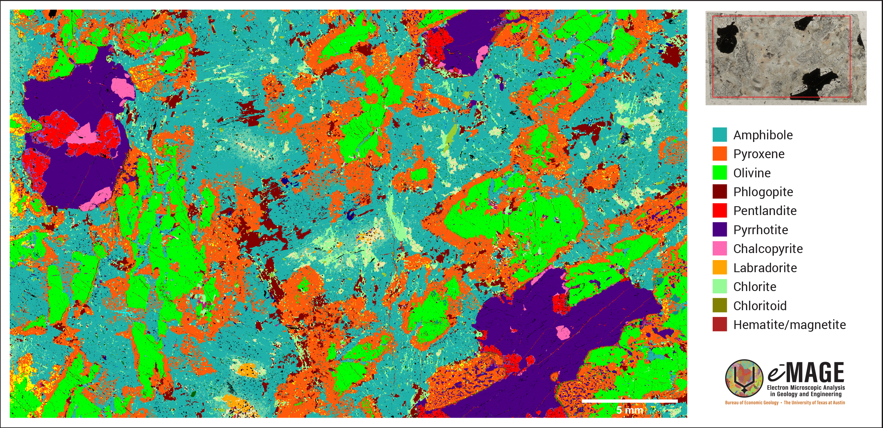
SEM-CL data combined with advanced Elemental Dispersive X-ray Spectroscopy (EDS) provides quantitative diagenetic and microstructural information that can be used for model input or validation.
e-MAGE Sample Preparation Equipment
- Zeiss-Axio Camera Imager.A2m automated stage Optical Microscope
- 2 Argon-Ion millers
- 2 Carbon coaters
- Iridium coater
- Saw, Polishing wheel, Sonicator, Oven
e-MAGE Lab ArcGIS SEM Petrography Database
The BEG e-MAGE Lab has created an ArcGIS-based SEM petrography database designed to make high-value imaging and quantitative petrographic data publicly visible, reusable, and collaboration-ready for internal and external interest groups and users. This is a multi-year initiative to capture, QC, and summarize legacy archives, with Phase 1 available here: https://arcg.is/1faOje3
The current platform is a prototype containing ~10% of our archive data. It is enough to demonstrate the structure, search power, and workflow while we continue scaling toward a comprehensive database and eventually building links to all raw images.
At its core, the system is a structured, searchable catalog of SEM-related datasets organized by formation, depth, geologic age, and data type. Users can filter and browse by:
(1) Formation / stratigraphic unit and basin/play context
(2) Depth and sampling interval
(3) Geologic age / stratigraphic framework
(4) Data type: SEM-SE, BSE, EDS maps, CL, AMICS/QEMSCAN-style mineral maps, experiments, derived products
(5) Image coverage: fields of view, mosaics, map extents, resolutions
(6) Associated publications and reports
Beyond imagery, the database supports quantitative petrography using standardized workflows (e.g., point counting, pore tracing, image-based measurements). All the metadata is validated and confirmed using S&P Global.
EPS E-beam Lab

The Department of Earth and Planetary Sciences (EPS) E-beam Laboratory specializes in spectroscopic analyses of mineral phases and major element compositions.
Equipment includes:
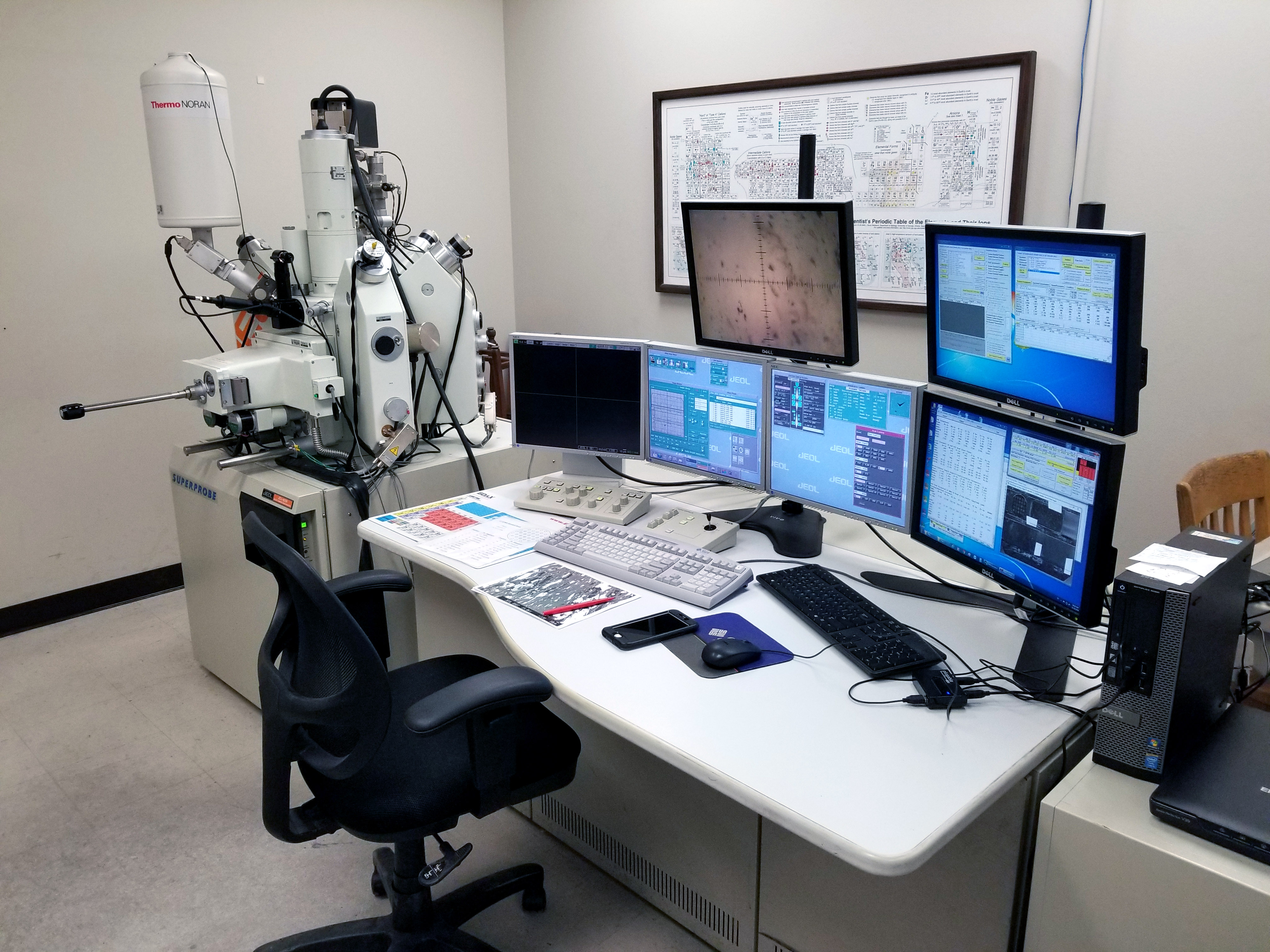
- JEOL JXA-8200 Electron Microprobe (EPMA) – Equipped with 5 wavelength dispersive spectrometers (WDS), secondary electron (SE), and backscattered electron (BSE) detectors.
- JEOL 6490 Low-Vac Scanning Electron Microscope – Equipped with electron-backscatter diffraction (EBSD), secondary electron (SE), backscattered electron (BSE), cathodoluminescence (CL), and energy-dispersive X-ray (EDS) detectors.
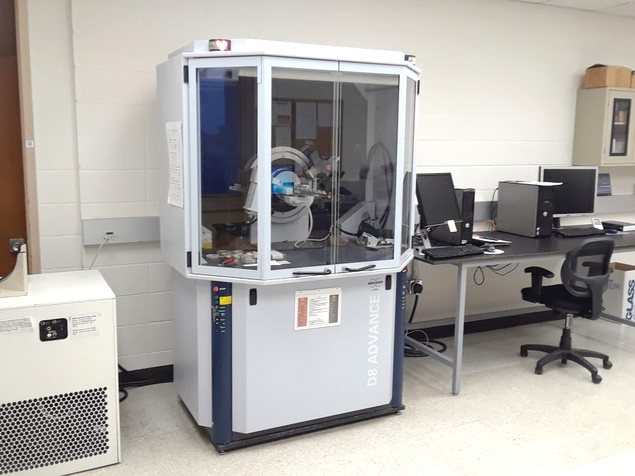
- Bruker D8 Advance Powder X-ray Diffractometer (XRD) – Provides routine, qualitative mineral identification and semi-quantitative modal mineralogy in rock powders.
- Raman Spectrometer
- Fourier Transform Infrared Spectrometer (FTIR)
TMI Electron Microscopy Facility

The Texas Materials Institute EM facility provides state-of-the-art sample preparation and atomic scale characterization instrumentation including TEM, FIB:SEM, SEM, STEM, and ESEM. The facility consists of a suite of specially designed low-vibration rooms with precise temperature and humidity control. Operation of TEM is done remotely in a separate room to remove the impact of people on the room stability. An array of specialized holders and detectors are available for state of the art in-situ capabilities and imaging.
Equipment includes:
- JEOL NEOARM Low kV STEM Corrected – Low-voltage, probe-corrected transmission electron microscope with aberration correction from 30 kV to 200 kV, equipped with atomic-level elemental mapping using EDXS and EELS, energy filtered imaging, electron binding state and plasma mapping, and in situ liquid cell, heating, and biasing.
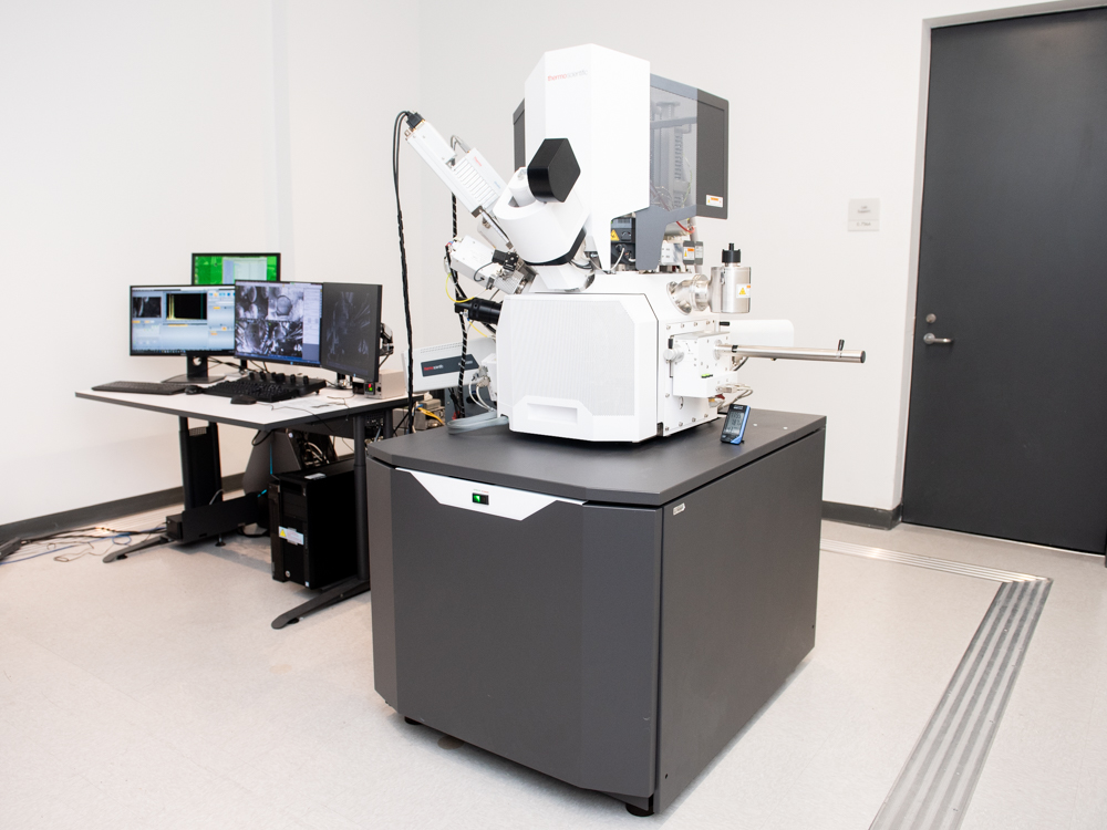
- Thermo Fisher Scios 2 HiVac Dual beam FIB:SEM – High quality TEM sample preparation with Sidewinder HT FIB, Easylift, and AutoTEM, subsurface and 3D characterization, high contrast UHR imaging with in-lense Trinity detection scheme, automated serial milling and imaging with Auto Slice and View 4.
- FEI Quanta 650 ESEM – Three imaging modes: high-vacuum, low-vacuum, and environmental SEM (ESEM) which enables charge-free imaging and microanalysis of non-conductive specimens without preparation. Bruker EDX for chemical analysis and Quorum Cyro-stage for specimen imaging in “natural” hydrated state.
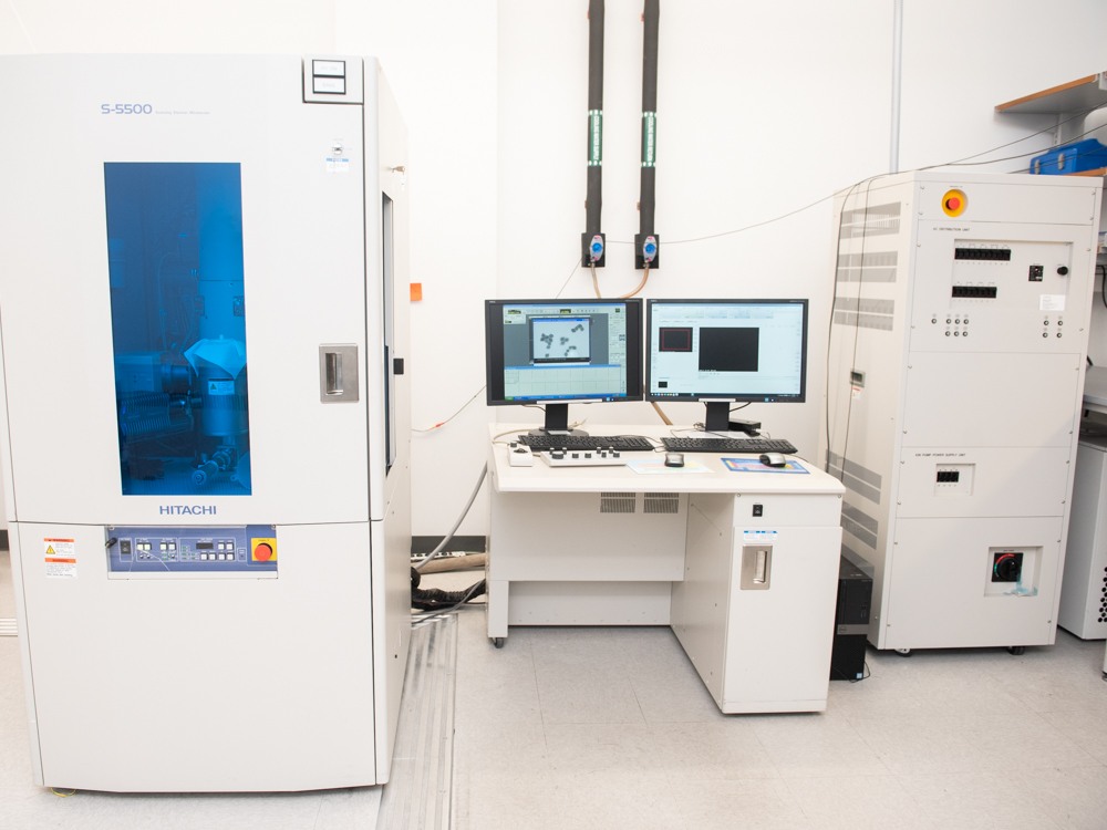
- Hitachi S-5500 SEM/STEM – Capable of scanning transmission electron microscopy (STEM) mode at sub-nanometer resolution, Duo-STEM detector for simultaneous observation of bright field (BF) and dark field (DF) STEM images, equipped with energy dispersive spectroscopy (EDS) providing chemical composition of materials at nanometer scale.
- Thermo Fisher Apreo 2C LoVac SEM – Low-vacuum imaging for charge-free imaging and analysis of non-conductive, outgassing, or beam-sensitive samples without metal coating.
Sample Preparation Equipment includes:
- Hitachi Ozone Cleaner
- EMS Sputter Coater
- Leica Cryo Setup
- Leica EM TIC 3X